Introduction:
The equine sarcoid is probably the commonest cutaneous tumour
in horses. All 6 forms of the disease have a high propensity
for recurrence and become more aggressive if subject to accidental
or iatrogenic interference.
The disease affects all breeds of horse, mules,
donkeys and zebra. There is some evidence that the thinner-skinned
breeds such as the Arabian have a particular tendency towards
the condition. Furthermore there is known to be a genetic basis
for the disease. Several genetic lines have known predisposition
but individuals within those lines may not get sarcoids at all
while others may be severely affected. While this may superficially
suggest that there is some heritable aspect of the disease it
is very important to realize that there are other factors that
need to be present for a particular animal to get the disease.
It is easy to propose that horses with sarcoids should not be
used for breeding but the genetic tendency to the disease probably
exists in a far higher number of horses than actually show overt
sarcoid skin disease. The suggested autosomal recessive gene
responsible for imparting a susceptibility to the condition
influences the severity and recurrence of the disorder in an
individual.
Realistically therefore we are not in any position
at this time to advise that affected horses should not be used
for breeding but I think it is reasonable to try to exert breeding
pressure against the disease by avoiding the breeding of two
affected horses.
Notwithstanding the genetic susceptibility,
I believe that no horse can be considered to be totally exempt
from the condition. Sex (geldings more commonly) and age (1-6
years) predilection have been proposed but recent studies suggest
that there is no significant breed, sex or age predisposition.
Sarcoids commonly multiply on the individual
horse; sometimes very rapidly while some others remain relatively,
or even completely, static for years. Interestingly, a few
individuals show spontaneous full and permanent self-cure.
Interestingly, in my experience, spontaneous full remission
(self-cure) usually means that the horse will not develop further
lesions. In a few cases treatment of one lesion (or a few)
has resulted in clinical improvement in others at other sites.
However, the course of the condition is entirely unpredictable
and it is probably unwise to assume that there any invariable
rules about the disorder: even the most benign-looking small
lesion can erupt into a potentially catastrophic mass in a short
time.
What causes the disease?
For many years researchers have been trying
to find a cause for the disease but we are still some way from
a definitive answer. The role of papilloma viruses is uncertain
- no patent virus particle has yet been conclusively demonstrated
but a very high proportion of sarcoids have genetic material
that is identical or very similar to that found in some papilloma
viruses. The distribution of lesions and the epidemiology of
sarcoids strongly suggest that flies are significant but how
the fly and the virus are linked is another matter yet to be
established.
What does the condition look like?
Clinically and pathologically, sarcoids present
most of the features of a true neoplasm; indeed I believe that
it is best regarded as a form of skin cancer. Although this
may not be strictly true in pathological terms it does at least
suggest that the behaviour of the tumour is unpredictable and
that treatment may be problematical. It is however clear that
it is not a wart!
Six distinct clinical entities which are noticeably
different can be recognized. Although each of these forms is
commonly identifiable it is important to recognise that the
“less severe” forms can rapidly progress to the more aggressive
types particularly if they are traumatised. Furthermore the
specific types may not be clearly identifiable in every case.
It is however, patently obvious that even the mildest forms
are indeed sarcoid - in vitro cell cultures derived from
these are typical and indistinguishable from those taken from
the more aggressive lesions. These factors suggest that both
cell and host factors are responsible in combination for the
variety of forms.
1. Occult Sarcoid
The predilection sites include the skin around
the mouth and eyes, the neck and other relatively hairless areas
of the body including the inside of the forearm, armpit and
thigh. Lesions show as hairless areas, often roughly circular.
They usually contain one or more small cutaneous nodules (2-5
mm diameter) or roughened areas with a mild hyperkeratotic appearance
but these may or may not be present or obvious in every case.
An area of changed/altered, slightly thickened skin with thin
hair coat and slight changes in hair pigment may be encountered
and may be difficult to identify in winter-coated animals.
The lesions are characteristically slow growing; they may progress
to “warty” verrucous growths or if injured may develop rapidly
into fibroblastic lesions. While the lesion remains as a static/quiescent
hairless patch showing no evidence of growth in size or number
of nodules, it may be wise not to interfere. Cases have existed
for over 15 years without treatment or acceleration; however
extensive development of verrucose sarcoid or conversion into
fibroblastic type sarcoid, usually demand immediate attention.
This can occur at any time with or without apparent insult.
2. Verrucous (Warty) Sarciod:
This type has a warty appearance with variable
degrees of flaking and scaling over limited or wider areas of
the body. Most often this type is seen on the face, body and
groin/sheath areas. Extensive areas can be affected and are
often surrounded by an area of slightly thickened /changed skin
(possibly reflecting a surrounding area of early occult sarcoid)
with altered, thin hair-growth pattern.
Individual lesions may be sessile (flat-based)
or pedunculated (with a narrow neck) giving a true wart like
appearance - indeed this type is probably the source of the
name “wart” on horses. The name is of course misleadingly benign
for a potentially dangerous condition. The lesions are most
often slow growing and not very aggressive until injured/insulted.
However, small nodules may appear at any stage or over any area
of the affected skin. These may develop a true fibroblastic
character whether or not they are insulted or traumatised. Rubbing,
biopsy, partial excision or minor or major trauma to the surface
commonly results in a dramatic change to fibroblastic sarcoid
over variable areas of the lesion.
The verrucose sarcoid can be mistaken for papillomatosis
(true warts), chronic blistering, severe chronic rubbing or
irritation such as can be seen in a few cases of sweet itch),
3. Nodular sarcoid:
The lesions are easily recognisable, as firm,
well-defined subcutaneous, spherical nodules of 5-20 mm diameter
but can be much larger. Most often this type can be found in
the groin, sheath or eyelid areas. The number of nodules varies
widely - single, few, several or hundreds are common. The nodules
usually lie under apparently normal skin and then may be freely
movable. However, sometimes there are dermal and deep attachments,
which prevent independent movement of the overlying skin and
/or movement of the tumour mass relative to deeper tissue.
The overlying skin may become thin over larger nodules and when
these ulcerate they quickly become more aggressive fibroblastic
type tumors. A similar aggressive fibroblastic response commonly
follows iatrogenic or accidental or iatrogenic damage.
4. Fibroblastic sarcoid:
These tumours have a characteristic fleshy appearance
and this type is commonly referred to as “Angleberries” (a name
that should be used only for a visually similar type of skin
tumour in cattle). Predilection sites include the groin,
eyelid, lower limbs and coronet, sites of skin wounds at any
location and sites of any other types of sarcoid subjected to
trauma or insult. Both pedunculated and extensive sessile tumours
with prominent ulceration and serum exudation are commonly encountered.
The latter may reflect single or repeated insults to the “lesser”
forms but may develop spontaneously. They are common at sites
of wounds (especially if other sarcoids are present elsewhere).
Accidental wounds that fail to heal may contain significant
sarcoid components in the wound margins and admixed with granulation
tissue. Surgical wounds are also liable to sarcoid development.
Failure of a surgical wound to heal in a horse with sarcoids
elsewhere could be associated with sarcoid transformation at
the site although there are many other possible causes of the
same problem. Concurrent excessive granulation tissue growth
serves to confuse the diagnosis.
In spite of their aggressive appearance they
do not metastasize but can spread locally in dermis by local
invasion/extension. Repeated insult (accidental or iatrogenic)
encourages local subdermal and dermal invasion.
This type of sarcoid looks very like “proud
Flesh” especially when it develops at the site of a wound and
more particularly at the site of limb wounds).
5. Mixed (Verrucous, Nodular and
Fibroblastic) Sarcoid:
This type of sarcoid probably represents a progressive/transient
state between the verrucous / occult types and fibroblastic
/ nodular types. Variations in proportion of the several types
of sarcoid is infinite and complex mixtures of any or all of
the above types (containing both verrucous and fibroblastic
elements) are common in long standing lesions or those subjected
to repeated minor trauma (such as rubbing by tack or harness).
They become progressively more aggressive as more fibroblastic
transformation takes place - a common consequence of biopsy
or injury.
6. Malevolent sarcoid:
This is a recently described variation with
predilection sites in the jaw, face, elbow and medial thigh
areas in particular. A particularly dangerous form occurs in
the immediate area around the eye. A history of repeated trauma
to other types of sarcoid e.g. surgical interference is commonly
described. Some cases have no such history with spontaneous
development of typical multiple, locally invasive sarcoids.
Others show extensive infiltration of lymphatics (cords of tumour
are commonly palpable) with numerous ulcerative nodules and
surface involvement as well as possible extension to local lymph
nodes.
The malevolent form of sarcoid is particularly
dangerous, not least because there is no current treatment for
it. Its appearance is not easily mistaken for other skin diseases
but again the presence of several different types of sarcoid
elsewhere on the body makes the diagnosis relatively simple.
Sarcoids generally have a high capacity for
local tissue invasion into the surrounding skin and other tissues.
This is particularly dangerous in the eyelid. This local spread
makes treatment very difficult and may explain why sarcoids
have a bad reputation for recurrences and the development of
new tumours following surgical excision or other interference.
One of the most dangerous problems that occur
with the sarcoid relates to those that develop at sites of wounds.
Even a small wound on the distal limb can become a very troublesome
sarcoid with a complete or partial failure of the wound to heal.
While the clinical appearance of proud flesh can be remarkably
similar, treatments for the two conditions are very different.
Indeed treatment that is suitable for proud flesh (cutting back
and grafting) serve only to make the sarcoid even more aggressive
and even more impossible to treat effectively. Thus, wound
management in all horses, and those with sarcoid skin tumours
at other sites (and those with no sarcoids that are genetically
susceptible to the disease) are particularly important.
So how can we be sure that a particular
skin lesion is, in fact, a sarcoid?
An individual lesion on a horse can be difficult
to diagnose although with experience most can be recognised.
The large majority of affected horses have more than one lesion
and many have over 25 – 30. Multiple tumors with characteristic
features of the various types of sarcoid on an individual horse
make the diagnosis simple - there are no other diseases with
the same range of clinical features and types. A few cases
are difficult nevertheless and then biopsy is sometimes required.
Some vets are understandably reluctant to interfere with a sarcoid
and so may not elect to obtain a biopsy. I have much sympathy
with this approach because this interference may trigger a massive
and uncontrollable expansion of the lesion. Particular difficulty
with diagnosis can arise when sarcoid is diffusely mixed with
granulation tissue. Biopsy may be very misleading if only one
of the two tissue types is recognized in the specimen. In most
cases diagnosis does not require a biopsy and so treatment can
be instituted immediately.
What treatments are there and how effective
are they?
Treatment should follow as soon after diagnosis
as possible. Suspicious lesions can justifiably be treated
immediately after biopsy using a suitable regimen. There are
ten or more recognised treatment methods for the disease and
this suggests that no one treatment is invariably effective.
The prognosis is always very guarded and owners
should be aware of the possible serious complications, which
can arise both from the disease itself and from the treatment.
The disease is best regarded as a form of skin cancer. Owners
must be aware of the limitations, cost and likely/possible outcome
of the various treatment options. There is a strong likelihood
that prolonged or repeated treatments will be required. We
are all looking for a “sure-fire” treatment for cancers but
this is a long way off yet for the equine sarcoid. No case of
sarcoid can be considered to be free of the disease even following
apparently successful treatment.
What factors will the vet consider before
selecting a treatment option?
The best option may be economically or practically
impossible. For example radiation carries a good prognosis
but is very restricted, requiring special conditions. Cryosurgery
of multiple lesions may require prolonged general anesthesia.
Very extensive lesions are virtually impossible to treat and
so an early attempt is justified.
Treatment Methods :
Many treatment methods have been used with varying
success. The various treatment methods described below may
be more or less applicable to specific types of sarcoid. Treatment
must remove every single abnormal cell - leaving even one behind
will inevitably, sooner or later, result in return of the tumour
(often with a more aggressive form).
Currently there is no effective treatment for
the malevolent form of the disease.
Ligation: A ligature of nylon thread,
a rubber band (or even tail hair) may be used around the base
of the lesion to cut off its blood supply. This method is not
feasible for flat or extensive lesions and those where the margins
of the sarcoid cannot be accurately defined.
Surgical excision: There is a high rate
of recurrence in all except the most confined and defined lesions
following surgical excision. Superficial (occult and verrucose)
lesions can be effectively treated by wide excision provided
that the wound can be closed and then protected during healing.
Any delay in healing may be due to sarcoid regrowth. Complete
or partial failure of the wound to heal within days of surgery
is a common indicator of problems but sarcoid regrowth can take
up to 5 or more years to recur at the site.
Notwithstanding the limitations of surgery,
excision of nodular lesions carries a somewhat better prognosis
provided that the procedure is performed correctly. Nodular
lesions in the eyelids however are potentially very dangerous
- they commonly have extensive ramifications through adjacent
tissues. Surgery often fails in these cases but there are some
success stories.
Fibroblastic, mixed and malevolent sarcoids
are generally not suitable for surgical excision alone. The
prognosis following surgery can be improved somewhat by combining
it with other modalities such as cryosurgery, topical cytotoxic
compounds, intralesional cisplatin injections or radiation
Cryosurgery (Freezing): Cryosurgery
is commonly employed. While some veterinary surgeons have good
success rates it has relatively poor overall success rates (except
in the smallest and most defined lesions, which carry a reasonable
success rate). Again, the whole lesion must be destroyed without
any significant damage to underlying or adjacent tissues. Local
scarring may have important effects on function, for example
of the upper eyelid or over joints.
Laser surgery (CO2-YAG laser
excision): Laser excision has a relatively high success
rate but again selection of the most appropriate lesions is
very important. The cosmetic results are however not often
acceptable. Equipment is expensive and is not commonly available.
Cytotoxic compounds: These induce extensive
tissue necrosis and scarring. They are easy to apply and relatively
cheap. Some complex mixtures of these with antimitotic, corticosteroid
and cytotoxic drugs have a reasonable reputation.
Cisplatin: This is a new method that
has only recently become available in UK. Good results are
reported for small fibroblastic and nodular lesions in particular
but it requires repeated injection into the lesion itself.
In some cases it can be used in conjunction with surgical debulking.
The material itself is very dangerous to humans and so its use
should be considered carefully.
Vaccines: Vaccines made from pieces
of the tumours have been widely used but the overall results
are singularly poor; it sometimes makes the condition even worse
class=MsoFootnoteReference> style='vertical-align:baseline;vertical-align:baseline'>.
If it worked to any acceptable degree it would surely have become
universal practice because it is a theoretically attractive
option for a virus disease. As a few cases of sarcoid skin
disease do resolve spontaneously it does imply that there may
be some immunological effects that might be used. The problem
is that we do not know what these are and furthermore we do
not know how to encourage this response. It is clear that vaccines
are not the most appropriate way of inducing this response.
Immunomodulation: Proteins including
various types of protein cell-wall fractions derived from Bacillus
Calmette-Geurin (BCG) have been used widely for sarcoid treatment
for many years. The material is injected repeatedly into the
individual sarcoids. Interestingly this method works best with
nodular lesions around the eye but away from the immediate peri-ocular
region and in other types of sarcoid there is less obvious benefit.
Some fibroblastic lesions on the limbs may in fact become worse
when treated in this way. The method is not appropriate for
mixed, verrucose or occult lesions for technical reasons. A
side issue of this method is the occurrence of very alarming
(and possibly even fatal allergic reactions) that may occur
within minutes or hours of injection. Fortunately we are aware
of how to minimise this risk but nevertheless it is always there.
Radiation: Radiation is the by far
the most successful treatment. However, it is extremely expensive
and very limited in availability. It is usually only used for
smaller lesions in areas for which no other method is suitable
such as the eyelids and over joints etc. If this treatment
is offered owners need to be made aware of the problems that
include extreme hazard to the surgeon who has to perform the
procedure. Rightly, few veterinary surgeons are willing to sacrifice
their own lives for a horse. In reality this treatment method
is best regarded as a last resort to be used when there is no
alternative.
Homeopathic remedies are often used to treat
sarcoids but in my experience are very disappointing. I think
it unwise to say that none of these will treat cases and so
I keep an open mind. Certain natural medicines including Allo
Vera, Rosemary Oil and Teetree Oil have however, been found
to help a few cases. It is also important to know that in some
cases application of remedies of various natural and homeopathic
types have resulted in considerable exacerbation of the tumours.
This is probably more a property of the fact that the tumours
have been interfered with rather than any directly harmful effect
of the remedy.
Summary:
The equine sarcoid is a cutaneous tumour only.
It does not spread to internal organs and so affected horses
may be perfectly normal in every other respect. However it
is also clear from my experience that individual animals can
be affected in less obvious ways. Some horses with few or large
numbers of lesions have improved dramatically in their behaviour
and performance when the lesions have been successfully treated.
This implies that there may be factors associated with the condition
that do indeed affect the function of other organs. As the
cells do not spread into the major organs we must assume that
there are chemical products that get access to the circulation
and that these may sometimes have metabolic consequences.
Whenever a horse is found to have a sarcoid
lesion it needs to be put into the proper perspective. If you
are buying a horse you need to know that the condition is unpredictable.
Before a purchaser parts with their money he/she should be sure
of the insurance implications and the likelihood that treatment
will be required. A single small lesion may remain identical
until the horse dies of old age but it could erupt at any time
or it may herald the development of more lesions as time passes.
It is clear that the fewer lesions that are present at any one
time the fewer it will get. I regard it as very important that
horses should be as sarcoid free as they can be over the summer
months when flies are a problem. Almost every owner of a horse
with sarcoids will recognise that the flies seem to congregate
at the site of sarcoids. Fly worry can sometimes cause bleeding
and severe worry to the horse.
Treatment at an early stage when there are few
small sarcoids is in my opinion the best approach. The prospects
for successful treatment are far better if the lesions are small,
early and the horse is under 4-5 years of age. None of the treatment
methods are cheap and none of them are certain of success –
if we can resolve 50% of lesions we are doing exceptionally
well. No matter how identical two lesions may appear to be,
the response to treatment can be very different - no two cases
respond in an identical fashion to a single treatment method.
Sarcoids around the face and on the legs are particularly dangerous
in almost every aspect of the disease and owners should not
be unduly surprised when any selected treatment fails to help:
indeed you should not be surprised if the treatment makes matters
worse! Furthermore there is no current method for treating microscopic
lesions – we can only treat those we can actually see. We would
hope of course that eventually we might be able to find a way
of making the immune processes of the patient recognise the
presence of abnormal cells and reject them – this way every
single cell could be detected and destroyed; there would be
no more sarcoids. This is some way off yet – if it were as
simple as this then we would have an answer to every cancer
and disease in every species of animal! We are in desperate
need of more effective treatments if we are to rid the horse
of this distressing and expensive disease.
The equine sarcoid should be regarded as a
form of skin cancer and should be treated seriously in every
case; early consultation with your vet will help to ensure that
the condition is held in check and not allowed to run rampant
through the skin of your horse.
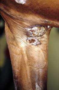
Figure 1:Mixed sarcoid in the
armpit of a bay gelding.
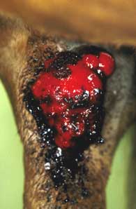
Figure 2: Severe fibroblastic
sarcoid on the medial (inside) thigh.
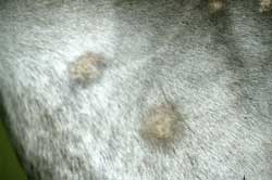
Figure 3: Occult lesion on the
thigh, note that they tie over large blood vessels.
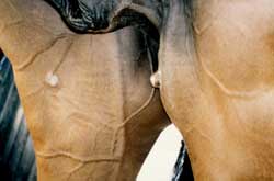
Figure 4: 2 small sarcoids, one
on blood vessel and a nodule on the other thigh.
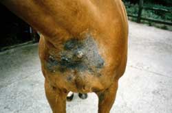
Figure 5: Extensive occult sarcoid
on the breast.
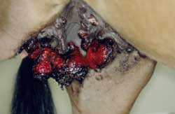
Figure 6: Malignant sarcoid in the groin of
a gelding. This eruption developed over 3 months from a relatively
minor group of small skin nodules. While we were moderately
successful in treating the case it was an expensive, persistent
and ongoing problem for the owner and the horse.
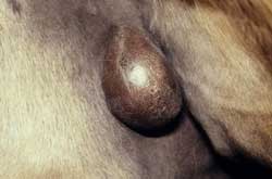
Figure 7: Single nodular sarcoid
in the thigh/groin.
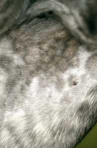
Figure 8: Extensive occult lesion
looking very like ringworm.
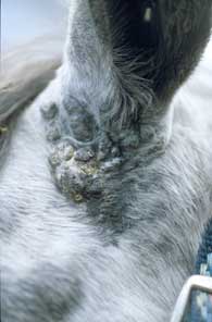
Figure 9: Mixed sarcoid around
the ear base. This is very difficult to treat.
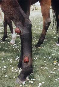
Figure 10: Sarcoids (Fibroblastic)
on two wounds that failed to heal.
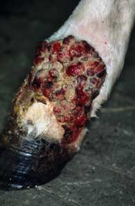
Figure 11: Severe sarcoid at the site of a small,
insignificant wound, resulted in the horse being destroyed.
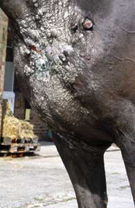
Figure 12: Extensive mixed sarcoid with verrucous
and nodular lesions. Some have ulcerated.
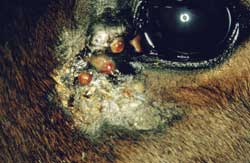
Figure 13: Very dangerous occular sarcoid that received radiation
treatment, which was very expensive but successful.
Knottenbelt DC, Edwards SER and Daniel EA
(1995) The diagnosis and treatment of the equine sarcoid In
Practice (supplement to Veterinary Record)